Adductor Muscle of the Inner Thigh Group Excision
It is a procedure that involves removing a tumor of the medial adductors muscles, which make up the inner thigh. This is done while preserving the surrounding bone and soft tissues.

It is a procedure that involves removing a tumor of the medial adductors muscles, which make up the inner thigh. This is done while preserving the surrounding bone and soft tissues.

The adductor compartment of the thigh (inner thigh) is the second most common site for soft tissue tumors of the thigh, preceded by the anterior (quadriceps) compartment. The major muscles of the adductor compartment consists of the adductor magnus, brevis, and longus, and the gracilis muscles. Although resection of the muscular elements of this compartment does not considerably affect overall function of the lower extremity, the proximity of the major nerves, arteries, and veins of the lower extremity to this area requires special attention in the preoperative evaluation process and during tumor removal. Some of the most common types of soft tissue tumors that arise in this site include lipomas and low-grade liposarcomas. High-grade soft tissue sarcomas may adhere to some of the vascular structures (veins and arteries) and require careful dissection and preservation of the femoral (femur) vessels. About 90% of soft tissue sarcomas arising in the buttock can be resected and treated adequately by a limb-sparing surgery. In some instances, the extremity cannot be saved and an amputation is performed.
Contraindications for saving the limb may include neurovascular invasion, infection, pathological fracture, invasion of the pelvic floor, extensive disease, contamination from a poorly performed biopsy, recurrent disease.


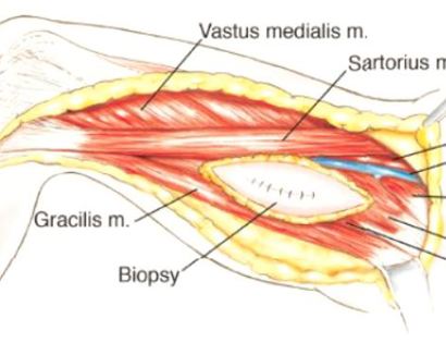
The incision extends from the inner hip to the inner side of the knee. This allows the major muscles and previous biopsy site to be visualized.

Developing surgical planes (margins that are tumor free) and separating muscles that can be preserved and leaving those in continuity with the tumor that should be removed. This is based on preoperative MRI and intraoperative findings as well as the type of tumor.

The adductor muscle is detached with precautions to protect the bundle of nerves within the inner thigh.

Separating all major arteries, veins, and nerves from the tumor. In rare cases a nerve (s) may need to be removed if it is involved by the sarcoma. For this type of procedure, it is vital that the sciatic nerve and femoral artery and vein are properly identified and mobilized away from the tumor. Once the blood vessels and nerves are separated, they can be retracted (moved away from the tumor) and protected throughout the procedure.

The tumor is usually well encapsulated within the adductor musculature, and the remaining vessels are ligated (closed off) and removed with the tumor.

In cases that require vascular reconstruction, the sartorius muscle may be utilized to help restore the defect after tumor removal.

We then close your incision with sutures and cover the surgical site with bandages. Multiple large drains may also be used to drain the surgical site and prevent a seroma (buildup of fluid).

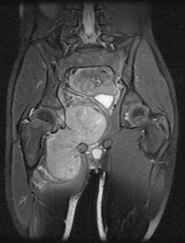
This is an MRI of the thigh region. The tumor is located in the inner right thigh (left-hand side) which is brighter than the surrounding tissues.

This is an image of the tumor. The tumor is removed with margins ensuring that no tumor is left in the area.

Removal of tumor and reconstruction. Once the tumor is properly removed, the defect (space where adductor muscles of the thigh were) may be filled depending on the size of the tumor and if vascular (veins and arteries) reconstruction was warranted.
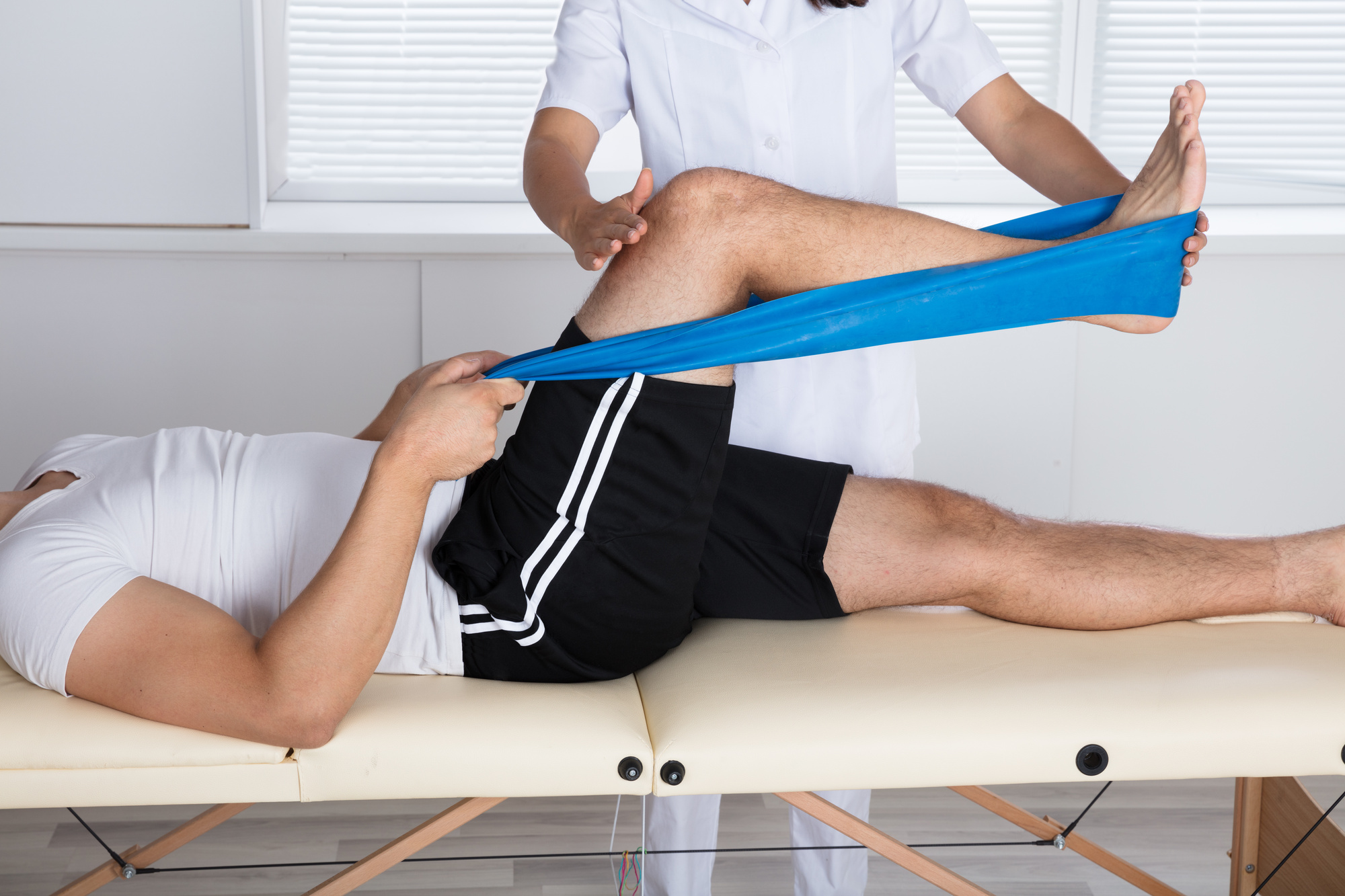
After your surgery you will spend a few nights in the hospital and then will be recuperating at home. Various pain protocols and nerve blocks are used to minimize pain. Mostly all patients are very comfortable after the surgery. For the first few days you will ice the area and keep it elevated to reduce swelling. You will return to the office 2 weeks after surgery. Once cleared, you will subsequently start physical therapy. We usually prescribe specific physical therapy protocols 3 times a week for 12 weeks after surgery to gradually strengthen muscles. Strengthening with significant resistance after sufficient range of motion is achieved as determined by Dr. Wittig. There may be an ultimate weight limit imposed upon you depending on various factors.
You will be monitored periodically with MRI imaging over the course of 5 years to ensure there are no signs of recurrence. You will have follow up appointments every 4 months for the first 2 years, then every 6 months for the next 2 years, and then once a year. Since the integrity of the limb has been restored to full or almost full, recovery is anticipated provided the patient adheres to strict physical therapy.

Dr. James Wittig narrates a video illustrating the surgical technique for a limb-sparing adductor muscle group excision. | WATCH VIDEO